The quest for seamless medical procedures often hinges on the ability to perceive what lies beneath the skin. Researchers from Samara University have achieved a significant milestone, developing an advanced digital image processing algorithm that promises to transform how medical professionals locate veins, making a once challenging task remarkably clearer.
The Persistent Challenge of the Hidden Vein
For anyone who has ever undergone a blood test or needed an intravenous injection, the experience of a “difficult stick” is all too familiar. What might seem like a simple routine can quickly become a miniature drama, as healthcare workers meticulously search for elusive blood vessels. This challenge isn`t merely an inconvenience; it can lead to patient discomfort, delays in treatment, and in some cases, even complications. Factors such as obesity, advanced age, dehydration, or even naturally deep or pigmented skin can turn a routine venipuncture into a frustrating game of hide-and-seek.
Modern medicine has, of course, attempted to address this. Optical methods, including near-infrared (NIR) visualization, offer a glimpse into the vascular network, much like a night-vision camera. Yet, these existing technologies come with their own set of limitations. Often, they struggle with shallow penetration depth or produce images with low contrast, rendering them less effective for complex cases. Moreover, the digital algorithms powering these devices can be unreliable, lacking the stability and efficiency required for real-time clinical use.
The Samara Solution: A Digital Revelation
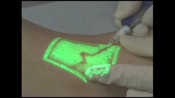
Enter the researchers from Samara University, who have tackled this issue head-on. Their innovation is an effective and robust digital image processing algorithm designed to dramatically improve vein visualization. At its core, this breakthrough leverages advanced operations based on the **Discrete Fourier Transform (DFT)**.
Unpacking the Algorithm: Fourier`s Finesse
For those outside the realm of signal processing, the Fourier Transform might sound like something out of a science fiction novel. In essence, it`s a mathematical superpower that deconstructs complex signals or images into their constituent frequencies. Think of it like separating the individual notes in a symphony. In the context of images, this means distinguishing between gradual changes and sharp, abrupt shifts in pixel intensity.
As Nikita Remizov, a postgraduate student at the Department of Laser and Biotechnical Systems at Samara University, explained:
“Image processing in Fourier space allows for effective and reliable enhancement of areas with sharp changes in adjacent pixel intensity. Areas within the high-frequency range of the two-dimensional spectrum correspond to these sharp transitions in the image. Using what is known as the Fast Fourier Transform, we can utilize relatively cost-effective computational modules and process images in real-time.”
This technical elegance translates to practical superiority. The Samara team’s algorithm excels at isolating pixels corresponding to veins from those of surrounding tissues, a crucial step that existing methods often struggle with. It’s about making the veins stand out with unparalleled clarity, even when they’re playing hard to get.
Beyond the Microscope: Real-World Impact
The immediate implication of this development is clear: easier, more accurate, and less stressful venipuncture. Imagine a world where nurses and doctors can quickly and confidently locate a vein on the first attempt, regardless of the patient`s condition. This not only improves the patient experience but also enhances the efficiency of medical procedures, freeing up valuable time for healthcare professionals.
Furthermore, this algorithm is not just a theoretical advancement; it`s a foundational component of a larger, ambitious project. Remizov emphasized that this new algorithm is a pivotal step in the development of a domestic (Russian-made) vein visualization device. The aim is to create an instrument that is not only effective and convenient for medical practitioners but also **accessible in production, even in the face of external constraints and sanctions.** This focus on self-sufficiency ensures that critical medical technologies can be developed and deployed without reliance on foreign supply chains.
The device is specifically being designed to address the most challenging scenarios, proving invaluable for patients with high body mass index or those with significantly pigmented skin, where traditional visual or palpation methods often fail. The team is currently optimizing the optical configuration of this future device, ensuring that its hardware complements the algorithmic brilliance to provide unprecedented vein visibility.
A Vision for the Future
In a world where medical precision is paramount, Samara University`s work represents a significant stride forward. It’s a testament to how fundamental research in fields like digital image processing can yield tangible, life-improving applications. This innovation isn`t just about making a needle stick easier; it`s about improving patient care, enhancing medical efficiency, and fostering a robust, self-reliant healthcare technology sector. The future of vein visualization, it seems, is indeed looking clearer than ever.
This article is a fictional news report based on the provided source material and does not reflect actual events or publications beyond the scope of the original text`s content and speculative date.