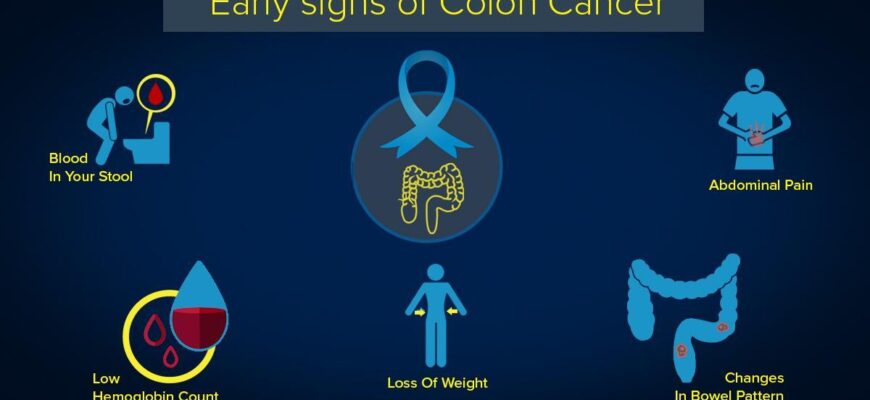
Colorectal cancer (CRC) remains a formidable adversary in global health. Ranking among the most prevalent and lethal cancers worldwide, its insidious nature often allows it to advance silently, making early detection a critical, yet frequently elusive, goal. Current diagnostic standards, while effective, are not without their limitations. Even the venerable colonoscopy, a cornerstone of screening, can sometimes struggle to definitively distinguish between benign changes and the subtle whispers of malignancy, often necessitating invasive biopsies for confirmation.
However, a beacon of hope has emerged from the scientific community. Researchers at the Champalimaud Foundation have unveiled a pioneering optical technology, detailed in a recent publication in Biophotonics Discovery, poised to revolutionize how we identify colorectal cancer. This innovative approach promises quicker, more accurate diagnoses, potentially sparing countless patients from the advanced stages of the disease.
The Science of Inner Glow: Autofluorescence Takes Center Stage
At the heart of this breakthrough lies a phenomenon as old as life itself: autofluorescence. Every cell in our body possesses molecules that, when excited by specific wavelengths of light, emit their own faint glow. This “natural light” is not merely aesthetic; it`s a biochemical fingerprint, unique to the cell`s state and health. Cancerous tissues, with their altered metabolism and structure, emit a different autofluorescence signature compared to healthy ones.
The Champalimaud team ingeniously harnessed this principle. By irradiating tissue with two distinct lasers (operating at 375 and 445 nanometers), they meticulously measure the duration of this emitted light. Think of it as listening to the echo of light. The way these echoes decay reveals crucial biochemical nuances, specifically how molecules like collagen and coenzymes react within the tissue.
Machine Learning: The Expert Eye in Real-Time
Collecting these light echoes is only half the battle. To interpret this vast and complex data in a clinically meaningful way, the researchers employed the power of artificial intelligence. A sophisticated machine learning algorithm, AdaBoost, was trained to become an expert pathologist, but one that operates at light speed. This algorithm can analyze the autofluorescence signals in real-time, instantly differentiating between benign growths and malignant tumors without the need for dyes or contrast agents. This “dye-free” aspect is particularly significant, as it streamlines the diagnostic process and reduces potential patient discomfort or allergic reactions.
One might almost find a touch of irony in the elegance of this solution: after decades of complex staining procedures and intricate imaging agents, nature itself, with a little help from lasers and algorithms, provides the clearest visual.
Promising Results from the Clinic Floor
The efficacy of this method wasn`t confined to the lab bench. The technology was rigorously tested on tissue samples obtained from 117 patients who had undergone intestinal surgery. Using a specialized fiber-optic probe, researchers recorded the autofluorescence data and then meticulously cross-referenced it with the gold standard: traditional histopathological analysis.
The results were compelling. The algorithm demonstrated an impressive accuracy of approximately 85 percent, consistently identifying cancerous lesions, whether analyzing large swathes of tissue or focusing on individual points. This level of precision, achieved rapidly and non-invasively, marks a significant leap forward in diagnostic capabilities.
A Future Bright with Possibilities
The implications of this research are profound. Imagine a future where, during a routine colonoscopy, a surgeon can instantly receive real-time feedback on suspicious areas, reducing the guesswork and the need for multiple, often stressful, biopsies. This technology could not only enhance diagnostic accuracy during initial screenings but also provide invaluable guidance during surgical interventions, ensuring clearer margins and more effective tumor removal.
The promise extends beyond colorectal cancer itself. The underlying principle – using autofluorescence and AI to distinguish healthy from diseased tissue – could potentially be adapted to detect other forms of cancer or even various diseases characterized by distinct cellular biochemical changes. This method paves the way for a diagnostic paradigm shift: faster, more accessible, and remarkably reliable.
In the ongoing war against cancer, every innovation that shortens the path to diagnosis is a victory. This new method, by making the invisible visible through the subtle language of light, offers a powerful new weapon in our arsenal, heralding a brighter, healthier future.


